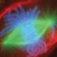
Fluorescence-stained Cell Undergoing Mitosis
Dublin Core
Description
A lung cell from a newt, commonly studied for its similarity to human lung cells, is stained with fluorescent dyes. The green stain reveals mitotic spindles, red is the cell membrane and part of the cytoplasm, and the structures that appear light blue are chromosomes. This cell is in anaphase of mitosis.
Contributor
Cut Rita Zahara
Rights
Creative Commons
Type
Files
Collection
Citation
“Fluorescence-stained Cell Undergoing Mitosis,” Open Educational Resources (OER) , accessed February 16, 2026, https://oer.uinsyahada.ac.id/items/show/943.


