Browse Items (29 total)
- Tags: Human Anatomy
Sort by:
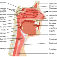
The Nose and its Adjacent Structures
Several bones that help form the walls of the nasal cavity have air-containing spaces called the paranasal sinuses, which serve to warm and humidify incoming air. Sinuses are lined with a mucosa. Each paranasal sinus is named for its associated bone:…
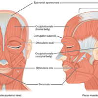
Muscles of Facial Expression
Many of the muscles of facial expression insert into the skin surrounding the eyelids, nose and mouth, producing facial expressions by moving the skin rather than bones.
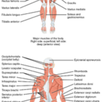
Overview of the Muscular System
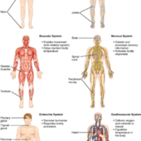
On the anterior and posterior views of the muscular system above, superficial muscles (those at the surface) are shown on the right side of the body while deep muscles (those underneath the superficial muscles) are shown on the left half of the body.…
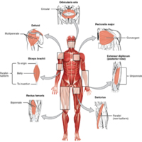
Muscle Shapes and Fiber Alignment
The skeletal muscles of the body typically come in seven different general shapes.
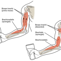
Prime Movers and Synergists
The biceps brachii flex the lower arm. The brachoradialis, in the forearm, and brachialis, located deep to the biceps in the upper arm, are both synergists that aid in this motion.
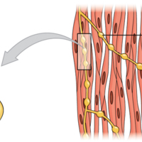
Motor Units
A series of axon-like swelling, called varicosities or “boutons,” from autonomic neurons form motor units through the smooth muscle.
Muscle Contraction
The dense bodies and intermediate filaments are networked through the sarcoplasm, which cause the muscle fiber to contract.
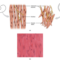
Smooth Muscle Tissue
Smooth muscle tissue is found around organs in the digestive, respiratory, reproductive tracts and the iris of the eye.
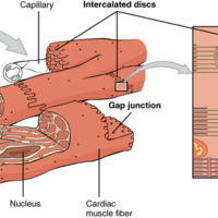
Cardiac Muscle
Intercalated discs are part of the cardiac muscle sarcolemma and they contain gap junctions and desmosomes.
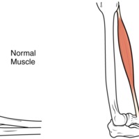
Atrophy
Muscle mass is reduced as muscles atrophy with disuse.
Tags: Anatomy & Physiology, Atrophy, biology, Human Anatomy
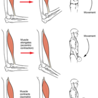
Types of Muscle Contractions
During isotonic contractions, muscle length changes to move a load. During isometric contractions, muscle length does not change because the load exceeds the tension the muscle can generate.
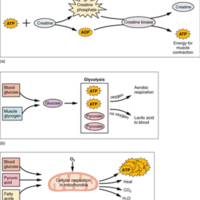
Muscle Metabolism
(a) Some ATP is stored in a resting muscle. As contraction starts, it is used up in seconds. More ATP is generated from creatine phosphate for about 15 seconds. (b) Each glucose molecule produces two ATP and two molecules of pyruvic acid, which can…
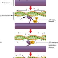
Skeletal Muscle Contraction
(a) The active site on actin is exposed as calcium binds to troponin. (b) The myosin head is attracted to actin, and myosin binds actin at its actin-binding site, forming the cross-bridge. (c) During the power stroke, the phosphate generated in the…
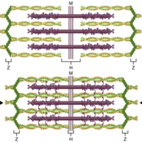
The Sliding Filament Model of Muscle Contraction
When a sarcomere contracts, the Z lines move closer together, and the I band becomes smaller. The A band stays the same width. At full contraction, the thin and thick filaments overlap completely.
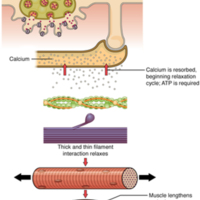
Relaxation of a Muscle Fiber
Ca++ ions are pumped back into the SR, which causes the tropomyosin to reshield the binding sites on the actin strands. A muscle may also stop contracting when it runs out of ATP and becomes fatigued.
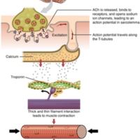
Contraction of a Muscle Fiber
A cross-bridge forms between actin and the myosin heads triggering contraction. As long as Ca++ ions remain in the sarcoplasm to bind to troponin, and as long as ATP is available, the muscle fiber will continue to shorten.
Human Anatomy and Physiology Preparatory Course
The overall purpose of this preparatory course textbook is to help students familiarize with some terms and some basic concepts they will find later in the Human Anatomy and Physiology I course. The organization and functioning of the human organism…
Tags: Human Anatomy, Physiology
Serous Membrane
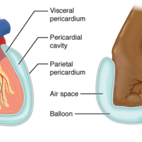
A serous membrane (also referred to a serosa) is one of the thin membranes that cover the walls and organs in the thoracic and abdominopelvic cavities. The parietal layers of the membranes line the walls of the body cavity (pariet- refers to a cavity…
Tags: Human Anatomy, Serous Membrane
Positive Feedback Loop
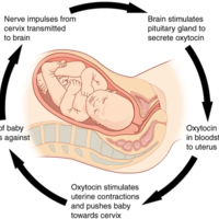
Normal childbirth is driven by a positive feedback loop. A positive feedback loop results in a change in the body’s status, rather than a return to homeostasis.
Levels of Structural Organization of the Human Body
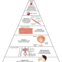
Before you begin to study the different structures and functions of the human body, it is helpful to consider its basic architecture; that is, how its smallest parts are assembled into larger structures. It is convenient to consider the structures of…
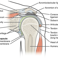
Shoulder Joint
The shoulder joint is called the glenohumeral joint. This is a ball-and-socket joint formed by the articulation between the head of the humerus and the glenoid cavity of the scapula ([link]). This joint has the largest range of motion of any joint in…
Tags: Human Anatomy, Shoulder Joint
Cardio
Cardio Exercises Below
1) Jog. You can do this outside on a treadmill or however you like.
2) Jump Rope routine 1
3) 10 Minute Jump Rope routine
4) Exercise Bikes
5) Sports Playing
1) Jog. You can do this outside on a treadmill or however you like.
2) Jump Rope routine 1
3) 10 Minute Jump Rope routine
4) Exercise Bikes
5) Sports Playing
Tags: Cardio, Human Anatomy
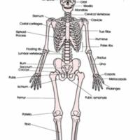
The Human Skeleton
The human skeleton provides shape and form to the human body
Our vital organs in our body are protected by our skeleton. More specifically our brain which is protected by what is called the skull and our heart and lungs are protected by our rib…
Our vital organs in our body are protected by our skeleton. More specifically our brain which is protected by what is called the skull and our heart and lungs are protected by our rib…
Tags: Human Anatomy, Human Skeleton
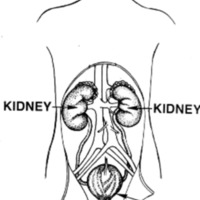
Excretory System
After food goes through the digestive system, the parts that are not digested need to be gotten rid of. That is the job of the excretory system.
Unabsorbed food goes to the large intestine. The liver also filters out solid particles of waste from…
Unabsorbed food goes to the large intestine. The liver also filters out solid particles of waste from…
Tags: Excretory System, Human Anatomy
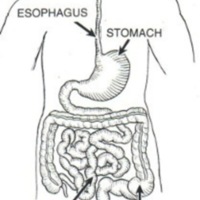
Digestive System
Our bodies need food to live and grow. The digestive system takes food and carries it to all the parts of the body.
The beginning of the digestive system is the mouth and teeth. Food that we eat has to be broken down into nutrients that cells in…
The beginning of the digestive system is the mouth and teeth. Food that we eat has to be broken down into nutrients that cells in…
Tags: Digestive System, Human Anatomy
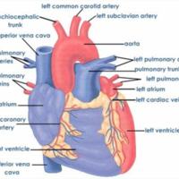
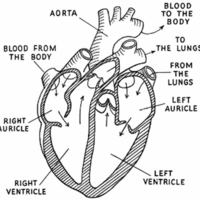
Circulatory System
The circulatory system includes the heart, blood, and a huge network of blood vessels that carry blood all over the body. The job of the circulatory system is to deliver oxygen to cells all over the body and then to carry out waste product like…
Tags: Circulatory System, Human Anatomy
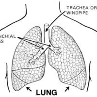
Respiratory System
The job of the respiratory system is to take oxygen from the air we breathe and get it to different parts of the body. Our bodies and the cells in them need oxygen (written with the chemical symbol O2) to live. Our cells give off carbon dioxide…
Tags: Human Anatomy, respiratory system


